Breakthrough in Breast Cancer Prevention: Scientists Target Cells-of-Origin in BRCA2 Carriers

Unveiling the Source of Cancer in High-Risk Individuals
In a landmark study led by the Walter and Eliza Hall Institute of Medical Research (WEHI) in Australia, scientists have made a significant breakthrough in the fight against BRCA2 gene breast cancer. The research, recently published in Nature Cell Biology, identifies for the first time the likely ‘cells-of-origin’. The specific cells that can develop into breast cancer in individuals carrying this genetic mutation.
Women inheriting the faulty BRCA2 gene face a daunting approximately 70% lifetime risk of developing breast cancer. Often at a younger age and with a tendency towards clinical aggression. Preventive strategies have thus far been limited to early screening and preventive surgeries, such as mastectomies, to lower this risk.
A Targeted Approach to Cancer Prevention
The study’s co-first author, Dr. Rachel Joyce, highlighted the discovery of an aberrant cell population within the breast tissue of BRCA2 carriers. These cells, identified in the majority of healthy tissue samples from carriers, divide more rapidly than normal. Thus suggesting their pivotal role as the originators of potential future breast cancers in these women.
Further insights from the study revealed these aberrant cells to be a subset of luminal progenitor cells in breast ductal tissues. Displaying altered protein production – a key factor in tissue growth and function. This alteration not only earmarks these cells as the likely origin of cancer. But also exposes a vulnerability to existing cancer treatments.
Towards a New Era of Prevention
Leading the research, Professor Jane Visvader and her team developed a pre-clinical BRCA2 model demonstrating similar alterations. They targeted these cells with Everolimus, an approved drug for relapsed breast cancer, showing that pre-treatment could delay tumor formation. This finding opens new avenues for preventive treatment strategies, aiming at specific aspects of protein production in these cells.
Despite the promising outcomes, the journey from lab to clinic remains cautious. Professor Geoff Lindeman, a study author and cancer clinician, emphasized the need for further research to refine these approaches, considering the potential side effects of drugs like Everolimus when used as preventive treatments. The goal is to develop more selective and tolerable treatments for BRCA2 mutation carriers, building on the significant strides already made in identifying the cells-of-origin for breast cancer linked to the BRCA1 gene mutation.
The study, supported by contributions from the Victorian Cancer Biobank, the National Health and Medical Research Council, and several other foundations, marks a critical step forward in the quest for effective breast cancer prevention strategies. It not only sheds light on the cellular beginnings of breast cancer in high-risk groups but also charts a course towards potentially life-saving treatments for women worldwide.
Stay informed and engaged with the latest advancements. Empower yourself with knowledge and make more informed decisions about your breast cancer treatment and care. Visit the Breast Advocate App website today and join us in the fight against breast cancer.
Breakthrough in Cancer Treatment: AI Algorithm Predicts Chemo Resistance
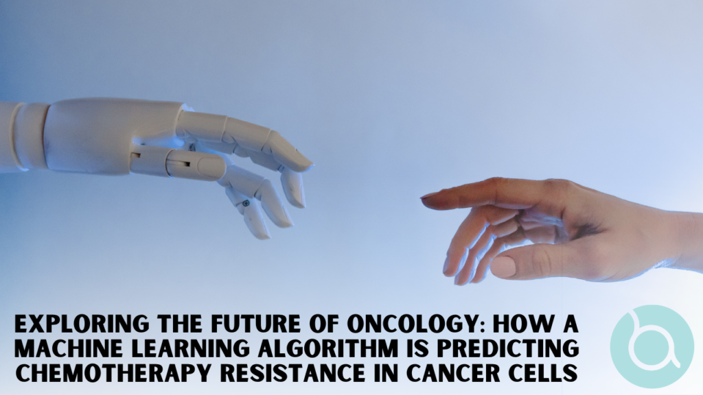
In an unprecedented leap for cancer research, scientists at the University of California San Diego School of Medicine have published a study in Cancer Discovery journal, unveiling a machine learning algorithm capable of predicting chemotherapy resistance in cancer cells. This ground-breaking development offers new hope in the fight against one of the deadliest diseases.
Tackling the Complexity of Cancer
Cancer cells, like all cells, depend on intricate mechanisms for DNA replication during cell division. Most chemotherapies target and disrupt this DNA replication in rapidly dividing tumor cells. However, predicting how a tumor will respond to such treatments has been a long-standing challenge in oncology.
This new algorithm, developed by UC San Diego researchers, addresses this issue by analyzing how a multitude of genetic mutations in a tumor collectively affect its response to DNA replication-inhibiting drugs.
Focusing on Cervical Cancer
The study specifically tested the model on cervical cancer tumors, using cisplatin, a widely used chemotherapy drug. Impressively, the algorithm successfully predicted treatment responses, identifying tumors most likely to resist treatment. It also shed light on the molecular mechanisms driving such resistance.
A Network-Based Approach
Trey Ideker, PhD, a professor in the Department of Medicine at UC San Diego, highlighted the model’s innovative approach. “Previous models focused on individual mutations, which lack significant predictive value. Our AI model analyzes thousands of mutations simultaneously, understanding the network-based nature of cancer,” Ideker explained.
The team focused on 718 genes commonly used in genetic testing for cancer, training their machine learning model with publicly available drug response data. This led to the identification of 41 molecular assemblies where genetic alterations impact drug efficacy.
Enhancing Treatment Predictions and Patient Outcomes
The AI model’s accuracy was evident in its application to cervical cancer, where approximately 35% of tumors persist post-treatment. It not only identified tumors susceptible to therapy, associated with improved patient outcomes, but also those likely to resist treatment.
Beyond Predictions: Understanding AI Decision-Making
What sets this model apart is its interpretability. Ideker emphasized the importance of understanding the AI’s decision-making process. “The transparency of our model is a key strength. It not only builds trust but also highlights new potential targets for chemotherapy,” he said.
Looking Forward
This breakthrough paves the way for enhancing current cancer treatments and developing new ones. The researchers remain optimistic about the broad applications of their model, hoping to revolutionize the approach to cancer treatment with this advanced AI-driven method.
Stay informed and engaged with the latest advancements. Empower yourself with knowledge and make more informed decisions about your breast cancer treatment and care. Visit the Breast Advocate App website today and join us in the fight against breast cancer.
Researchers Discover Breakthrough in Immunotherapy’s Side Effects
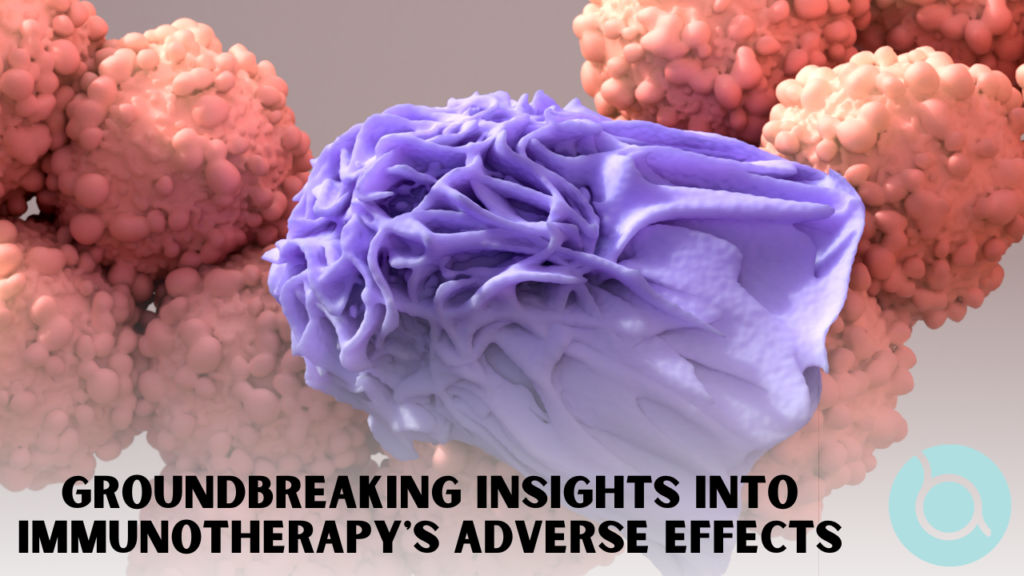
Researchers at the University of Michigan Health Rogel Cancer Center have made a significant breakthrough in understanding and mitigating severe gastrointestinal problems associated with immune-based cancer treatment. This discovery could lead to more effective cancer therapies with fewer adverse effects.
Immunotherapy’s Promise and Pitfalls
Immunotherapy has gained prominence as a promising approach for treating various types of cancer. However, one of its downsides is the potential for severe side effects, such as colitis. Colitis is inflammation in the digestive tract. Colitis can cause substantial discomfort and may even lead some patients to discontinue their cancer treatment.
The challenge for scientists was that while patients experienced colitis, laboratory mice did not. This made it difficult to study the underlying causes of this side effect. To address this, the research team, led by Bernard C. Lo, Ph.D., developed a new mouse model. They injected microbiota from wild-caught mice into the traditional mouse model, which then exhibited colitis symptoms after receiving immunotherapy antibodies.
Unlocking the Mechanism: The Role of Gut Microbiota
Through this innovative model, the researchers were able to identify the specific mechanism responsible for colitis. It was revealed that colitis resulted from the composition of the gut microbiota. This caused immune T cells to become hyperactive while simultaneously deleting regulatory T cells. Which typically inhibit T cell activation in the gut. Crucially, this effect was limited to a specific domain of the immune checkpoint antibodies.
Researchers removed that particular domain and found that it still elicited a robust anti-tumor response without inducing colitis. Dr. Gabriel Nunez, the senior study author, stated, “Once we identified the mechanism causing the colitis, we could then develop ways to overcome this problem and prevent colitis while preserving the anti-tumor effect.”
Collaboration and Future Endeavors: Moving Towards Clinical Trials
The antibody used to prevent colitis was developed by Takeda Pharmaceuticals. The Rogel team plans to conduct further studies to gain a deeper understanding of the mechanisms causing colitis. They are also actively seeking clinical partners to advance this knowledge into a clinical trial.
The study’s authors include Ilona Kryczek, Jiali Yu, Linda Vatan, Roberta Caruso, Masanori Matsumoto, Yosuke Sato, Michael H. Shaw, Naohiro Inohara, Yuying Xie, Yu Leo Lei, and Weiping Zou.
Funding for this research was provided by grants from the National Institutes of Health, Takeda Millennium Pharmaceuticals, the Canadian Institutes of Health, the Crohn’s and Colitis Foundation, and the National Science Foundation. The Rogel Cancer Center Shared Resources, including Single Cell Spatial Analysis, Tissue and Molecular Pathology, played a crucial role in supporting this work.
Stay informed and engaged with the latest advancements. Empower yourself with knowledge and make more informed decisions about your breast cancer treatment and care. Visit the Breast Advocate App website today and join us in the fight against breast cancer.
New Study Links Gut Microbiome to Breast Health

In a groundbreaking study, researchers have uncovered a potential link between the human gut microbiome and breast health. Shedding new light on how diet and lifestyle factors may influence breast cancer risk. The study focuses on the impact of flaxseed components known as lignans, and their interaction between gut microorganisms and the expression of mammary gland microRNAs (miRNAs). Some of which play a role in regulating genes associated with breast cancer.
Insights from Leading Researchers
Jennifer Auchtung, Ph.D., Assistant Professor in the Food Science and Technology Department at the University of Nebraska — Lincoln, emphasized the importance of this discovery. Stating, “The gastrointestinal microbiota plays an important role in modifying many components of our diet to impact human health. In this study, we found correlations between diets enriched in flaxseed, cecal microbiota composition, and miRNA profiles in the mammary gland that regulate many pathways. Including those involved in cancer development. This preliminary study supports further research into the role that the microbiota plays in dietary approaches to reduce risk factors associated with disease.”
Manipulating Microbiota-Mammary Gland Interactions
Investigating whether the relationship between gut microbiota and mammary gland miRNAs could reduce breast cancer risk. Researchers administered flaxseed lignan components to female mice. The cecum (a part of the colon located in the lower right abdomen) was of particular interest. It is believed to play a role in producing short-chain fatty acids and harboring anaerobic bacteria crucial for metabolism.
One of the lignans found in flaxseed oil requires microbial processing to release bioactive metabolites with potential antitumor effects. The study ultimately revealed a significant relationship between the microbiota and mammary gland miRNA, with flaxseed lignans modifying this relationship to be non-cancer-causing.
Elena M. Comelli, Ph.D., Associate Professor at the University of Toronto’s Department of Nutritional Sciences, Temerty Faculty of Medicine, and the corresponding author of the study, expressed optimism. She said, “If these findings are confirmed, the microbiota becomes a new target for preventing breast cancer through diet.”
This study opens the door to further research on gut microbiome in breast health and the potential for dietary interventions. Also, offering new hope for the development of preventive strategies in the fight against breast cancer.
Stay informed and engaged with the latest advancements. Empower yourself with knowledge and make more informed decisions about your breast cancer treatment and care. Visit the Breast Advocate App website today and join us in the fight against breast cancer.
AI Sparks Excitement: Reducing Chemotherapy for Breast Cancer Patients

Groundbreaking AI Tool Outperforms Expert Pathologists
A groundbreaking Artificial Intelligence (AI) tool could revolutionize breast cancer treatment. According to a recent study by Northwestern Medicine, AI could reduce the need for chemotherapy in some patients. The study, published in Nature Medicine, revealed that AI evaluations of patient tissues outperformed expert pathologists in predicting the future course of breast cancer. Potentially sparing patients from the side effects of unnecessary chemotherapy.
Breast cancer is a major health concern. Approximately 300,000 U.S. women are expected to receive a diagnosis of invasive breast cancer in 2023. With one in eight U.S. women likely to face a breast cancer diagnosis in their lifetime. The need for more precise and personalized treatment options is greater than ever.
The Role of Non-Cancerous Cells in Predicting Outcomes
Traditionally, pathologists have relied on the examination of cancerous tissue to determine the severity and treatment approach for breast cancer. However, the Northwestern Medicine study highlights the importance of assessing non-cancerous cells in predicting patient outcomes.
Professor Lee Cooper and the Northwestern University Feinberg School of Medicine research team have developed an advanced AI model. Analyzing both cancerous and non-cancerous elements in breast cancer tissue. It assesses 26 properties to generate overall and individual prognostic scores for cancer, immune, and stromal cells. This detailed analysis offers a clearer picture to pathologists and can assist in creating personalized treatment plans.
One of the most significant implications of this AI tool is the potential to reduce chemotherapy duration. Chemotherapy can come with unpleasant and harmful side effects, such as nausea or heart damage. The ability to tailor treatment more precisely is a welcome development.
Furthermore, the AI tool could allow for treatment adjustments based on real-time tissue analysis. This approach holds promise for improving patient outcomes and reducing disparities in breast cancer care. Great news for those diagnosed in community settings with limited access to specialized pathologists.
The study was made possible through collaboration with the American Cancer Society (ACS). The AI model was trained with the help of medical students and pathologists worldwide. Students contributed thousands of annotations of cells and tissue structures in digital images of patient tissues.
Validation and Future Developments
As Northwestern Medicine transitions to using digital images for diagnosis, the AI model’s integration into routine breast cancer assessment could become a reality soon.
In addition, the research team is developing AI models tailored to specific breast cancer types, including triple-negative or HER2-positive. Enhancing the accuracy of predictions and provide deeper insights into breast cancer biology.
Funding Support for Promising Research
This promising research is supported by grants from the National Cancer Institute of the U.S. National Institutes of Health, marking a significant step toward a more personalized and effective approach to breast cancer treatment.
Stay informed and engaged with the latest advancements. Empower yourself with knowledge and make more informed decisions about your breast cancer treatment and care. Visit the Breast Advocate App website today and join us in the fight against breast cancer.
New Research Reveals Potential Breakthrough in Fighting Breast Cancer Metastasis

In a groundbreaking study, scientists from Penn State have uncovered crucial insights into how breast cancer cells invade healthy tissues. Shedding light on potential new ways to combat the deadliest aspect of cancer: metastasis. This discovery has the potential to revolutionize cancer treatment and offers fresh hope for patients battling breast cancer.
The research, led by Penn State’s Erdem Tabdanov, assistant professor of pharmacology, and published in the journal Advanced Science. It marks a paradigm shift in our understanding of cancer cell motility. The study reveals that a motor protein known as dynein plays a pivotal role in powering the movement of cancer cells.
Erdem Tabdanov explained the significance of this discovery. He states, “Until now, dynein has never been caught in the business of providing the mechanical force for cancer cell motility, which is their ability to move themselves. Now we can see that if you target dynein, you could effectively stop motility of those cells. Therefore, stop metastatic dissemination.”
Collaboration Across Institutions
The study originated as a collaboration between Penn State’s Department of Chemical Engineering and Penn State’s College of Medicine. Eventually it expands into a multi-institution partnership. Including researchers at the University of Rochester Medical Center, Georgia Institute of Technology, Emory University, and the U.S. Food and Drug Administration.
To understand the mechanics of breast cancer cell movement, researchers utilized live microscopy. Helping to observe the migration of live breast cancer cells in two different systems modeled after the human body.
Dynein’s Role in Cancer Cell Movement
The first system, is a two-dimensional network of collagen fibers. It demonstrated how cancer cells move through the extracellular matrix, powered by dynein. Dynein was identified as a key player in this process, shedding light on its significance in cancer cell movement.
Amir Sheikhi, assistant professor of chemical engineering and biomedical engineering at Penn State, emphasized the importance of their findings. Sheikhi says, “Instead of killing the cancer cells with radiation or chemotherapy, we are showing how to paralyze them. This is great news because you don’t really have to kill the cells. […] A harsh approach that targets both cancerous and healthy cells. Instead, you just have to stop the cancer cells from moving.”
Promising Implications for Patient Treatment
Tabdanov added that this “cell paralysis” approach could prove to be a more effective treatment strategy for cancer. Particularly after the surgical removal of the tumor, it could prevent cancer from spreading without causing harm to healthy tissues.
Compared to traditional chemotherapy, which aims to eliminate cancer cells at a faster rate than the rest of the body, this innovative approach focuses on containment rather than destruction. “The trick with chemotherapy is to kill the cancer cells slightly faster than the rest of the body — it’s a race against time,” Tabdanov noted. “Chemotherapy causes a lot of damage to the body’s normal, healthy tissues while it is busy killing the cancer. If we instead contained the cancer, stopped it in its tracks, we could keep the healthy parts of the body healthy.”
This groundbreaking research offers new hope in the fight against breast cancer, metastasis and provides a promising avenue for the development of more targeted treatments. Further studies and clinical trials will be needed to translate these findings into practical applications. However this discovery represents a significant step forward in the battle against breast cancer.
By staying informed and engaged with the latest advancements, you can empower yourself with knowledge and make more informed decisions about your breast cancer treatment and care. Visit the Breast Advocate App website today and join us in the fight against breast cancer. Together, we can work towards a brighter and healthier future for all those affected by this challenging disease.
3D Modelling Technique Helps Understand Complex Breast Cancers Better

3D Modelling of Complex Breast Cancers
Researchers at the University of Waterloo have developed a new way to better understand complex cancers using advanced 3D modelling techniques. This breakthrough might change how doctors approach treatment, especially for breast cancer.
What’s the big deal?
- In the past, scientists studied cancer by growing the cells in flat dishes. This method, however, didn’t fully represent how the cancer looks and behaves inside the body.
- Now, with a combination of modern bioprinting and microfluidic chips (tiny structures that imitate the flow of blood around a tumour), researchers can create a more accurate 3D model of the cancer. This means they can study it in a way that’s closer to how it really is in the body.
Why is this especially important for breast cancer patients?
Breast cancer is tricky. When it spreads, it forms complex tumours with different types of cells. This new 3D model helps researchers understand these complex tumours much better. The old way, which relied on just a sample or two, sometimes missed the full picture, leading to treatments that might not work as well.
How did they do it?
- They made tiny structures (microfluidic chips) that act like the environment around a real tumour.
- They grew different cancer cells in a special mixture (called bioink) that keeps them alive.
- Using a machine similar to a 3D printer, they placed these cells onto the chips, creating a living 3D model of the cancer.
What’s the end goal?
This advanced 3D model can be used to test treatments, like chemotherapy drugs. It’s a step forward in finding more effective treatments for breast cancer and other complex cancers, potentially leading to better outcomes for patients.
In short, this research offers hope for improved breast cancer treatments and a deeper understanding of the disease. Every new piece of research brings hope and better understanding. This latest finding might be a step closer to ensuring more effective and prolonged treatments for patients. As always, we will keep our community updated on the latest and most relevant information.
Shared Decision-Making: How Patients Feel Predicts Treatment Success

The study showed that breast cancer patients’ self-perceptions better indicate treatment effectiveness than clinician tools. The research also underscores the crucial role of shared decision-making in treatment strategies.
The study found that clinicians sometimes overestimate patients’ physical health compared to patients’ self-reports.
The Essence of Shared Decision-Making:
“Shared decision-making, a cornerstone of patient-focused care, involves both the doctor and patient discussing available evidence on the pros and cons of treatment options. This ensures the most suitable and informed choices are made,” Lead author Natansh Modi, an NHMRC PhD candidate at Flinders University’s Clinical Cancer Epidemiology Lab, stated.
The clinician uses the ECOG PS tool, while patients use PROs to share their perspectives during the decision-making process.
Study Findings:
Published in ESMO Open, this research combined data from numerous trials, observing nearly 3000 patients treated for HER2-positive breast cancer.
Mr. Modi elucidated, “We deduced that several patient-reported outcomes, notably regarding physical and mental health, were pivotal in determining the patient’s survival rate, cancer progression, or severe side effects during treatment.”
This study suggests patient data predicts treatment outcomes better than clinician scores. “About 70% of patients deemed ‘fully active’ by their clinicians actually reported restrictions in their physical health upon self-evaluation,” Modi remarked.
Mr. Modi concluded, “This research indicates that patient-reported physical well-being surpasses clinician-evaluated ECOG PS in predicting prognosis. It’s crucial for clinical practice to evolve, placing more weight on the patient’s viewpoint.”
Future Implications:
Integrating patient surveys with clinician evaluations can elevate shared decision-making in cancer care and refine future clinical trial designs.
Given these findings, it’s crucial for breast cancer professionals to prioritize patient insights and shared decision-making in treatment. To truly harness the potential of a patient-centric approach, we urge professionals to visit tolimanhealth.com. The platform offers resources and tools specifically designed to facilitate the creation of personalized shared decision-making tools for patients. By integrating these tools, clinicians can better align treatments with patients’ real-time experiences, fostering improved outcomes and patient satisfaction.
New Findings Suggest a Potential Shift in the Fight Against Breast Cancer Treatment Resistance
Breaking Down the Latest Breast Cancer Treatment Research

Breast cancer cells can sometimes be sneaky. After treatment, a few might not die off completely, but instead “play dead” temporarily, only to bounce back later. Researchers puzzle over why some cells resist treatment. A recent study now offers some insight into this, suggesting a possible game-changer in how we approach breast cancer treatment, Unveiling Tariquidar.
What The Research Says:
- The Sneaky Cells: The study shows that some breast cancer cells enter a ‘safety mode’ after treatment.In this mode, they sort of “play dead” by temporarily halting their growth, changing some of their characteristics, and becoming more resistant to drugs. This is known as a “drug tolerant persister” (DTP) state.
- The Role of P-gp: The protein called P-glycoprotein (P-gp) plays an essential role in this process.When the cancer cells are in this DTP state, they pump up the levels of P-gp, which acts like a safety shield. This protein helps remove harmful molecules produced by drugs like doxorubicin.
- A Potential Solution: The exciting news is that there might be a way to tackle these sneaky cells. The study shows that a drug called Tariquidar can block the action of P-gp. When used alongside chemotherapy, Tariquidar may prevent these cells from bouncing back. In fact, in experiments with mice, using Tariquidar increased their survival rate significantly.
What Does This Mean For Patients?
This discovery may lead to a shift in how we tackle breast cancer treatment resistance. Instead of always trying to fight the resistant cells, the focus might change to preventing them from becoming resistant in the first place.
Furthermore, adding Tariquidar to the treatment regime might offer a better outcome without causing more side effects, since it’s targeting the resistance mechanism directly.
In Conclusion:
Breast cancer treatment is a complex journey. Every new piece of research brings hope and better understanding. This latest finding might be a step closer to ensuring more effective and prolonged treatments for patients. As always, we will keep our community updated on the latest and most relevant information.
Air Pollution and Increased Risk of Breast Cancer – What You Need to Know

In a new study, researchers from the National Institutes of Health (NIH) have identified a concerning link between living in areas with high levels of particulate air pollution and increased cases of breast cancer. That is to say, this critical research aims to shed light on the impact of environmental factors on health.
Here’s what the findings mean for you:
About the Pollution
The pollution discussed is fine particulate matter. Specifically those particles 2.5 microns in diameter or smaller (PM2.5). These tiny particles, are from sources like vehicle exhaust, combustion processes, and industrial emissions. These can be inhaled deeply into our lungs.
Increased Risk
The study found that women living in areas with higher PM2.5 levels are more likely to develop breast cancer. Specifically, the risk went up by 8% for those in areas with higher air pollution. While this might seem like a small number, it’s significant considering how common air pollution exposure is.
How Can You Check Air Quality?
The Environmental Protection Agency offers a tool called Air Now. By simply entering your zip code, you can find out the air quality, including PM2.5 levels, in your vicinity.
Why This Study is Unique
What sets this research apart is its deep dive into historical air pollution levels. In contrast, many other studies focus only on current pollution levels. Furthermore, cancers, including breast cancer, can take years to develop. Therefore, this broader view may give a clearer understanding of how past air pollution affects current health outcomes.”
Different Types of Tumors
The study discovered that PM2.5 exposure is more linked with ER+ breast cancers. These are the most commonly diagnosed tumors in U.S. women. This hints at a biological process where the particulate matter disrupts our endocrine system.
What’s Next?
This study has offered vital insights. Undoubtedly, the researchers believe there’s more to learn! Especially regarding how different kinds of PM2.5 exposures might affect breast cancer risks in various regions.
The study’s findings emphasize the importance of reducing air pollution and being aware of our environment’s potential health impacts. If you’re concerned about your risk, it may be helpful to speak with a healthcare professional. Also, consider checking air quality in your area.
Stay connected with Breast Advocate to keep up to date with the lastest Breast Cancer News.