AI May More Accurately Predict Breast Cancer Risk
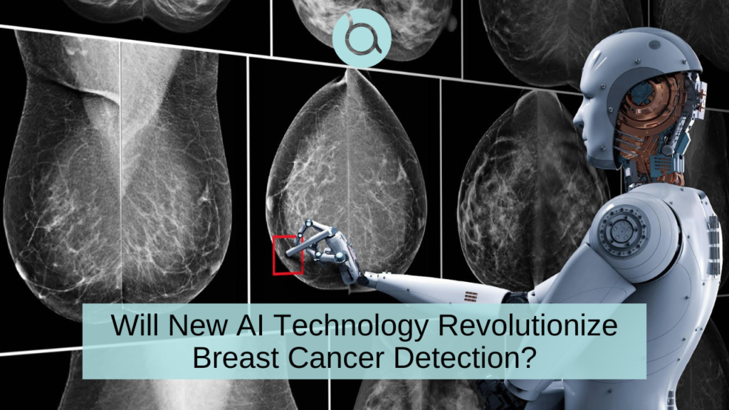
Early detection plays a crucial role in improving breast cancer survival rates and treatment outcomes. The integration of artificial intelligence (AI) into radiologic imaging, like mammograms and MRIs, is revolutionizing breast cancer diagnosis.
In a recent study, artificial intelligence (AI) outperformed the standard clinical model for predicting the five-year risk for developing breast cancer.
AI Study of Screening Mammograms
Lead investigator, Dr. Vignesh Arasu, used data from screening mammograms at Kaiser Permanente in Northern California in 2016 that showed no visible evidence of cancer. “We selected from the entire year of screening mammograms performed in 2016, so our study population is representative of communities in Northern California.”
Five different artificial intelligence (AI) algorithms were used to generate risk scores for developing breast cancer over the five-year period using the 2016 screening mammograms. The risk scores were then compared to the Breast Cancer Surveillance Consortium (BCSC) clinical risk score as well as to one another.
“All five AI algorithms performed better than the risk model for predicting breast cancer risk at 0 to 5 years,” Dr. Arasu said. “This strong predictive performance over the five-year period suggests AI is identifying both missed cancers and breast tissue features that help predict future cancer development. Something in mammograms allows us to track breast cancer risk. This is the ‘black box’ of AI.”
Advancements in AI for Breast Cancer Diagnosis
AI technology has made significant strides in recent years, bringing a paradigm shift in the field of breast cancer detection.
- Mammograms: AI-based systems can analyze mammographic images and assist radiologists in detecting early-stage abnormalities.
- Breast MRI: AI algorithms can analyze MRI images to identify suspicious areas, helping radiologists pinpoint potential cancerous lesions more accurately. By combining the power of AI with radiologic testing, medical professionals can achieve comprehensive and reliable breast cancer diagnosis.
Benefits of using AI
The integration of AI in breast cancer diagnosis offers numerous benefits:
- AI algorithms can assist in reducing false negatives and false positives, which are a persistent challenge in conventional mammography.
- AI can significantly enhance the efficiency and speed of breast cancer detection.
- AI can help bridge the expertise gap in breast cancer diagnosis.
Challenges and Ethical Considerations
While AI brings tremendous promise, it also raises important challenges and ethical considerations.
One critical challenge is the need for robust and diverse datasets to train AI algorithms effectively. Transparency and interpretability of AI algorithms are also crucial. Understanding the decision-making process of AI systems is vital for radiologists and patients to trust and validate the results.
We must also address issues surrounding patient privacy and data security. AI systems require access to extensive patient data, including medical records and imaging studies, which must be protected to safeguard patient confidentiality.
Striving for Better Patient Outcomes
AI algorithms can provide radiologists with powerful tools to improve accuracy, efficiency, and speed in diagnosing breast cancer lesions. By reducing false negatives and false positives, AI can also improve sensitivity and specificity, leading to better patient outcomes.
Will this detect breast cancer 5 yrs earlier?
MIT’s Computer Science and Artificial Intelligence Lab has developed a new tool that uses artificial intelligence (AI) and deep learning to predict the development of breast cancer up to five years earlier than current detection techniques.
Unlike other AI-based tools that use data mostly from acquired from white patients and may therefore be algorithmically biased, MIT’s model works equally well for both white and black patient populations. This is particularly important as black women are 42% more likely to die from breast cancer than white women – a possible contributing factor could be that current detection techniques don’t work as well in black women.

To create the tool, data was obtained from over 90,000 mammograms and outcomes from over 60,000 patients treated at the Massachusetts General Hospital. A form of machine learning known as “deep learning” was then used to identify patterns in the images and data that are too subtle to be routinely recognized by human physicians. The results have so far have been more accurate than current diagnostic approaches, presumably because the model is not based on existing knowledge and assumptions about patient risk factors.
MIT’s project is intended to help doctors compile the best personalized screening program for their patients and hopefully eliminate the heartbreaking outcome of a late breast cancer diagnosis. We eagerly await to hear if and when this could become widely available.